Bridging gap between AI Technology and Clinical practice
Medical Image Annotation is a foundational element in the Advancement of AI
Medical Image Annotation and Its Challenges
Key challenges in Medical Image Annotation
Variability
Medical images vary due to differences in equipment and techniques, requiring large datasets that are challenging to gather.
High Costs and Scalability
Reliance on experts like physicians leads to high costs and limited scalability in medical imaging annotation.
Data Privacy and Security
Maintaining a fine balance between giving annotators access and ensuring data security is crucial.
Manual Intensive
The need for expertise and the variability of images make the annotation process very manual-intensive.
Physician Burnout
The intensive manual annotation process puts a significant strain on medical professionals.
Specialize Knowledge
Requires deep medical expertise to accurately identify anatomical structures and abnormalities.
Caddie's solutions are for?
Al Companies
Several AI companies rely on accurate and expertly annotated medical data to develop robust algorithms. AI companies who want to accelerate research and gain regulatory compliance, Caddie’s medical image annotations are available in leading image data centers and CRO services powered by experts will help reach their goals faster.
University Researchers
Clinical Researchers
Clinical researchers are venturing into uncharted territories using AI and data analytics to unlock new possibilities in healthcare. Caddie’s AI tools not only offer operational efficiency but also provide Scientific AI Powered by generative AI (gen AI) and foundational models can revolutionize trial design and success rates.
Our Collaborators
Pioneering Medical AI Excellence: Visionary Minds Uniting to Transform Healthcare
Through Advanced Image Analysis and Innovative Artificial Intelligence Solutions

Dr. Kunio Doi
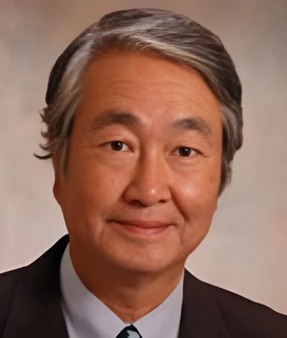
Emeritus Professor, Univ Chicago, Recognized as father of CAD using computerized image analysis and artificial intelligence, that improves diagnoses using clinical images worldwide

Dr. Maryellen Giger
A.N. Pritzker Distinguished Service Professor of Radiology, University of Chicago Member of US National Academy of Engineering (NAE)

Dr. Fleming Lure
Received the first FDA pre-market approved (PMA) early-stage lung cancer detection system on radiograph.

Dr. Stefan Jaeger
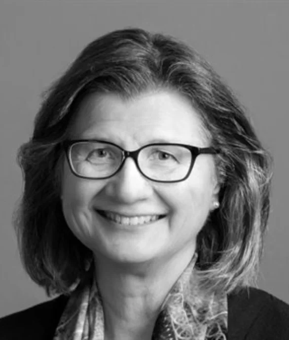
He is a staff scientist at the Lister Hill National Center for Biomedical Communications at the United States National Library of Medicine (NLM), which is part of the National Institutes of Health (NIH).

Vassilios Raptopoulos
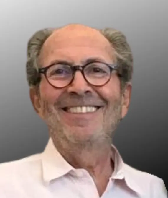
Professor of Radiology, Harvard Medical School, and Vice Chair for Clinical Services at Radiology Department of Beth Israel Deaconess Medical Center. Former President of the New England Roentgen Ray Society.
Industries We Serve
Healthcare
Auto
Manufacturing
Technology
Retail
Defence
Testimonials
High efficiency of SIFT predictions makes it attractive for tuberculosis diagnostics and monitoring. Multi-domain, patient-centric databases from NIAID TB Portals could be used to align SIFT predictions with retrospective data on treatment history, drug sensitivity, history of relapses, influence of comorbidities and treatment outcomes.
For AI to add the most value for patients and physicians, it needs to support, not supplant
MD, a clinical assistant professor of medicine at Stanford University
SIFT promises adoption of federated learning by offering a critical component in powering a sustainable, inclusive learning system.
Get in touch
Accelerate your research and enhance diagnostic accuracy with Caddie's solutions.
Connect with us
ADDRESS:
Ste 300 #1238
Gaithersburg, MD 20879
Please fill out the form below:
Our latests news
Nam molestie metus et justo hendrerit, non molestie velit imperdiet
vivamus ac sapien vitae dui mattis tristique cras finibus convallis ex vel lacinia justo
